Understanding Craniodiaphyseal Dysplasia: Causes, Symptoms, and Care
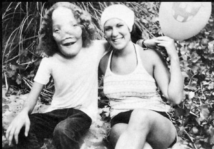
If you’re searching for information about craniodiaphyseal dysplasia, you’re likely seeking answers about a condition that’s incredibly rare and complex. Known by the nickname lionitis, craniodiaphyseal dysplasia (CDD) is a genetic bone disorder that leads to severe calcium buildup in the skull and facial bones, drastically changing facial structure and compressing the brain and cranial nerves. Although there are only a few documented cases worldwide, understanding this disease is essential for anyone affected or researching rare genetic conditions.
What Is Craniodiaphyseal Dysplasia (CDD)?
Craniodiaphyseal dysplasia is an extremely rare autosomal recessive disorder that causes excessive bone growth, particularly in the craniofacial region. The condition results in progressive thickening of the skull and facial bones, which not only affects appearance but also leads to neurological complications due to the compression of the brain and cranial nerves.
The overgrowth of bone narrows the openings in the skull, which can disrupt the function of the eyes, ears, and other vital nerves. This intense pressure often leads to vision problems, hearing loss, and sometimes even mental decline due to the restricted space for the brain.
Genetic Origins of Craniodiaphyseal Dysplasia
Craniodiaphyseal dysplasia follows an autosomal recessive inheritance pattern. This means that both parents must carry and pass on the defective gene for their child to be affected. Even then, the condition is extraordinarily rare, making it one of the least understood bone disorders in medical literature.
To date, no single gene mutation has been definitively identified as the cause of all CDD cases. However, researchers believe that abnormal bone remodeling — particularly involving calcium and phosphate regulation — plays a central role in the disease’s progression.
Symptoms and Clinical Features of CDD
The symptoms of craniodiaphyseal dysplasia are both physical and neurological, and they often become noticeable in early childhood. These include:
- Prominent facial features due to bone overgrowth (wide-set eyes, enlarged forehead, flattened nasal bridge)
- Macrocephaly (an abnormally large head)
- Severe headaches caused by increased intracranial pressure
- Vision loss due to pressure on the optic nerves
- Hearing impairment from compression of the auditory nerves
- Sinus obstruction and breathing difficulties
- Delayed cognitive development or decline
The bony thickening can be seen through X-rays, CT scans, or MRIs, which often show a “cloud-like” appearance due to the density and spread of the calcified bone tissue.
How Is Craniodiaphyseal Dysplasia Diagnosed?
Diagnosing craniodiaphyseal dysplasia involves a combination of clinical examination, imaging tests, and family medical history. Pediatricians often spot abnormal head growth or facial asymmetry early on, which may lead to further evaluation by specialists.
Key diagnostic tools include:
- Skull X-rays to observe bone density and formation
- CT or MRI scans for detailed imagery of cranial structures and nerve compression
- Genetic testing (where possible) to detect any mutations associated with bone remodeling
Due to its rarity, CDD is frequently misdiagnosed as other conditions like craniosynostosis, frontometaphyseal dysplasia, or osteopetrosis, which share some overlapping features.
Treatment Options and Management
There is no cure for craniodiaphyseal dysplasia, and treatment typically focuses on symptom management and improving the patient’s quality of life.
Surgical Interventions
Surgeries may be performed to relieve pressure on the brain and cranial nerves. These procedures include:
- Cranial decompression surgery
- Facial reconstruction
- Shunt placement to drain excess cerebrospinal fluid
Medications and Therapies
While medications cannot reverse the bone overgrowth, corticosteroids and vitamin D inhibitors have been used to attempt to slow progression. However, results are limited and not always consistent across cases.
Supportive Therapies
- Speech therapy
- Hearing aids
- Vision correction
- Occupational and physical therapy for motor development
Life Expectancy and Prognosis
Unfortunately, the prognosis for individuals with craniodiaphyseal dysplasia is often poor. Many children with severe forms of the disease experience life-threatening complications in early childhood due to increased intracranial pressure and respiratory issues. However, there have been rare cases of individuals surviving into adulthood with surgical intervention and continuous medical support.
It’s important to remember that each case is unique. With early diagnosis and a multidisciplinary care approach, some complications can be managed to enhance the individual’s quality of life.
The Emotional Impact and Need for Support
Living with craniodiaphyseal dysplasia affects more than just physical health. It can take a deep emotional toll on both the patient and their family. The noticeable facial differences, repeated surgeries, and developmental challenges often lead to social isolation and psychological distress.
Support groups and rare disease organizations can provide emotional support, advocacy, and access to clinical trials or experimental therapies. Connecting with other families facing similar challenges is invaluable for navigating the complex road of rare disease management.
Craniodiaphyseal Dysplasia vs Other Bone Disorders
To fully grasp the uniqueness of craniodiaphyseal dysplasia, it helps to compare it with similar conditions:
| Condition | Key Feature | Difference from CDD |
| Osteopetrosis | Bone overgrowth | Denser bones throughout the body, not just the skull |
| Craniosynostosis | Premature fusion of skull bones | Doesn’t involve widespread calcium buildup |
| Fibrous dysplasia | Bone replaced by fibrous tissue | Causes weak, deformable bones rather than excessive density |
| Frontometaphyseal dysplasia | Facial and skeletal anomalies | Less skull involvement, different genetic basis |
Raising Awareness and Research Needs
Because craniodiaphyseal dysplasia is so rare, there is limited funding and research devoted to understanding it. Most medical professionals never encounter a case in their lifetime, which makes awareness efforts even more crucial.
Promoting more studies into the genetic origins, bone metabolism pathways, and potential gene therapies is essential. Each new discovery can bring hope to affected families and potentially lead to breakthrough treatments.
How to Support a Loved One with CDD
If someone you know has been diagnosed with craniodiaphyseal dysplasia, your support can make a tremendous difference. Here are ways you can help:
- Be informed. Learn about the condition so you can understand the challenges involved.
- Offer emotional support. A listening ear is often more helpful than advice.
- Help coordinate care. Managing specialists, appointments, and therapies can be overwhelming.
- Join or support advocacy groups. These organizations work to improve funding and research.
Hope for the Future
Advances in genetics, biotechnology, and 3D imaging are bringing new hope for conditions like craniodiaphyseal dysplasia. While we still have a long way to go, each step forward adds to the understanding of this baffling condition.
By supporting awareness and advocating for further medical research, we contribute to a future where CDD can be better understood, treated, and perhaps even prevented.
Conclusion
Craniodiaphyseal dysplasia may be one of the rarest bone conditions in the world, but it deserves our attention, compassion, and scientific curiosity. With early diagnosis, coordinated care, and ongoing research, individuals affected by this challenging disorder can find support, relief, and hope.
Frequently Asked Questions (FAQs)
Q1. What is craniodiaphyseal dysplasia?
A: Craniodiaphyseal dysplasia (CDD) is a very rare genetic bone disorder that causes excessive calcium buildup in the skull and facial bones, leading to abnormal thickening and distinct facial features.
Q2. What are the main symptoms of craniodiaphyseal dysplasia?
A: Common symptoms include enlarged head size, wide-set eyes, severe facial deformities, narrowed nasal passages, and in some cases, neurological complications due to compressed brain structures.
Q3. What causes craniodiaphyseal dysplasia?
A: CDD is caused by a mutation in an autosomal recessive gene, meaning a child must inherit the mutated gene from both parents to develop the condition.
Q4. Is there a cure for craniodiaphyseal dysplasia?
A: There is currently no cure for CDD. Treatment focuses on managing symptoms, improving quality of life, and performing surgeries to relieve pressure on the brain and nerves.
Q5. How is craniodiaphyseal dysplasia diagnosed?
A: Diagnosis involves imaging tests like X-rays and CT scans to examine bone thickening, along with genetic testing to confirm the specific mutation.
Q6. How rare is craniodiaphyseal dysplasia?
A: It is extremely rare, with fewer than 30 documented cases worldwide. Because of its rarity, research and treatment options are still limited.
Keep an eye for more latest news & updates on Hamro Solarllc!